mide-kanseri

Stomach Cancer

Stomach cancer is the fourth most common type of cancer. It develops as a result of malignant tumors forming in the stomach lining due to various reasons such as poor diet, smoking, and genetic predisposition. More common in men, stomach cancer tends to metastasize to the lymph nodes, liver, and lungs. As with all types of cancer, early diagnosis makes treatment easier.

Causes of Stomach Cancer

Stomach cancer can develop and spread from any part of the digestive system. There are several risk factors for its development. These factors can be listed as follows:

  • Poor nutrition (Processed foods, excessive consumption of animal products, ready-made supermarket products, excessively salted vegetables create a زمینه for the development of stomach cancer.)
  • Infection (The most important factor is Helicobacter pylori infection.)
  • Smoking
  • Alcohol
  • Genetic predisposition
  • Symptoms of Stomach Cancer
  • Indigestion
  • Bloating
  • Weight loss
  • Abdominal pain
  • Nausea
  • Vomiting
  • Feeling of fullness

The most important symptoms of stomach cancer are indigestion and bloating. In advanced stages, abdominal pain, nausea, vomiting, and weight loss may occur. Patients over 40 who have not previously experienced these complaints should pay attention to symptoms such as indigestion and weight loss.

Stomach Cancer Treatment

There are different types of stomach cancer. Treatment depends on these types. Cancers detected in the early stages make surgery easier. In surgery, the tumorous section or the entire stomach can be removed. In patients whose stomachs are removed, a new stomach is created from the intestine. These patients can lead normal lives. They only need to follow the prescribed diet.

Diseases

rektum-kanseri

Rectal Cancer

The 10-12 cm section at the end of the large intestine, just in front of the anus, is called the rectum. It is where stool is temporarily stored. When sufficient stool accumulation occurs, the rectum stretches, creating the sensation of defecation. The rectum consists of two parts: the upper and lower rectum. The lower part remains empty except during constipation.

Rectal cancer develops when polyps in this area transform into tumors. It is more common in men than women, especially men over 50, who are at the highest risk. However, the incidence increases after the age of 35. Genetic predisposition is important in rectal cancer. If someone in the family has had rectal cancer, the risk increases in other individuals.

Causes of Rectal Cancer

There are several risk factors for the development of rectal cancer, primarily genetic predisposition. These factors can be listed as follows:

  • Being male
  • Irregular and unhealthy diet (Especially excessive consumption of animal products and processed meat products)
  • Sedentary lifestyle (Insufficient physical activity)
  • Obesity (More common in overweight individuals)
  • Smoking
  • Genetic predisposition (If one family member has had rectal cancer, other family members are also at risk.)

Symptoms of Rectal Cancer

  • Recurrent bowel irregularities such as constipation and diarrhea,
  • Rectal bleeding (Blood in the stool),
  • Decreased stool diameter,
  • Difficulty defecating,
  • Pain

Rectal Cancer Treatment

Rectal cancer can be treated with surgery, radiotherapy, and chemotherapy. Surgery may involve either complete removal of the rectum or removal of the cancerous area, depending on the stage and spread of the tumor. Surgery, radiotherapy, and chemotherapy should be applied in combination.

Diseases

laparoskopi

Laparoscopy

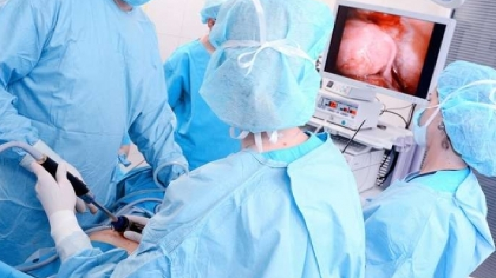
Laparoscopic surgery is a minimally invasive surgical method. Using thin, long instruments and a video camera, imaging and treatment are performed through small incisions of 0.5-1 cm. Laparoscopic surgery does not require large incisions like open surgery, which is its biggest advantage. Because the incisions are very small, the patient recovers more quickly, returns to normal life faster, does not experience pain after surgery as in open surgery, and the risk of complications is very low. The inclusion of a camera allows for a more detailed and comfortable surgical process. The camera images are transmitted to a television monitor, allowing the surgeon to observe even the finest details while simultaneously performing the treatment.

Advantages of Laparoscopic Surgery

  • Less pain is felt after surgery and it causes less trauma
  • The patient recovers faster
  • Earlier discharge
  • Faster return to normal life
  • Fewer wounds and more aesthetically pleasing scars
  • Less adhesions in the abdomen
  • Lower risk of complications.

Open Surgery

In open surgery, a large incision is made to visualize the abdominal organs. These incisions can be in different locations depending on the organs. They are usually 15-30 cm in size. This varies depending on the surgery and the patient’s size. The instruments used are those used in open surgery. After the surgery, the abdominal muscle membranes and skin are sutured separately. Manual manipulation of the internal organs, stretching of the abdominal wall with a retractor, and tight sutures cause significant pain and discomfort for the patient. It takes longer for the gastrointestinal system, in particular, to regain its normal function. All of this delays the patient’s hospital stay and return to normal life. The development of open surgery occurred after the discovery of anesthesia and antisepsis (in the 1850s). Therefore, its history spans approximately 165-170 years.

Laparoscopic surgery, on the other hand, was first used by gynecologists for simple procedures. Laparoscopic cholecystectomy (minimally invasive gallbladder surgery) became widespread after 1990. This was followed by colon surgery. It has been one of the most exciting advancements in the field of surgery in the last 25 years.

In Which Diseases is Laparoscopy Applied?

In the following cases, the laparoscopic method has moved beyond being experimental, developed, and become standard treatment:

Diseases

vucut-kitle-indeksi-hesaplama-formu

Body Mass Index Calculation

BMI Calculator
Use this calculator to check your body mass index (BMI).
BMI Calculator
Height
cm.
Please enter value between 125 and 225.
Weight
kg.
Please enter value between 10.0 and 500.0 ( one decimal ).
Height
ft.
in.
Please enter value between 4.2 and 7.3
Weight
lb.
st.
Please enter value between 22.0 and 1000.0 ( one decimal ).
Calculate BMI
Use this calculator to check your body mass index (BMI), which can be a helpful tool in determining your weight category. Or, use it to calculate your child’s BMI.

Diseases

Endoscopy

Endoscopy is a procedure for visually examining hollow organs. Endoscopy is a general term; the actual name of the procedure varies depending on the organ being examined. For example, examining the esophagus, stomach, and duodenum together is called gastroscopy. Endoscopy performed on the large intestine is called colonoscopy.

Endoscopy procedures are performed under sedation, a state of conscious sleep. The patient is conscious but in a semi-sleep state and therefore does not experience discomfort. They remember nothing after the procedure.

Gastroscopy

In gastroscopy, a lighted and flexible instrument is inserted through the mouth and can reach down to the duodenum. So, which diseases are diagnosed using gastroscopy?

  • In diseases affecting the esophagus (difficulty swallowing, burning and pain in the back of the chest, bitter taste in the mouth, etc.)
  • In diseases affecting the stomach and duodenum (burning, pain, heartburn in the upper abdomen, nausea, vomiting, anemia resistant to treatment, unexplained weight loss, etc.)
  • In upper digestive system bleeding (manifested as fresh blood coming from the mouth, vomiting resembling coffee grounds, or black stools)
  • In the diagnosis of esophageal, stomach, and duodenal cancers
  • In the detection of ulcers, inflamed areas, and small tumors
  • In widening strictures and removing polyps (fleshy protrusions) without surgery
  • In removing swallowed foreign objects
  • Gastroscopy is preferred for bleeding control.

Colonoscopy

Colonoscopy is used to diagnose and, if necessary, treat problems in the large intestine through the rectum. A flexible endoscope, longer than a gastroscopy, is inserted through the rectum. Diseases related to the large intestine are investigated by observing a distance of approximately 20-120 cm. In which cases is a colonoscopy used?

  • Bleeding in the stool,
  • Black stools
  • Anemia that does not respond to treatment
  • Changes in stool shape such as flattening
  • Unexplained weight loss
  • Unexplained abdominal pain
  • Colonoscopy is used in cases of prolonged diarrhea and constipation.
  • Colonoscopy is also important in the early diagnosis and treatment of intestinal pellets.
  • Individuals aged 50 and over who have no complaints should have a colonoscopy every 5 years. Because colon cancer is a treatable disease when diagnosed early.

What is Rectosigmoidoscopy?

Rectosigmoidoscopy is a shortened form of colonoscopy. It is a procedure that examines the last 60 cm of the large intestine. This section is called the sigmoid colon because of its resemblance to the letter “S”. Unlike colonoscopy, there is no need for preparation the day before the procedure. It is sufficient to cleanse the bowels with an enema in the hospital. It is a 5-minute procedure. It is recommended for young patients with typical hemorrhoidal or fissure (crack, tear) bleeding.

Diseases

apandisit

Appendicitis

The appendix, also known as the cecum, is a 6-9 cm long, tube-shaped organ located at the beginning of the large intestine. While its exact function is unknown, it is known to contain lymphatic tissue and produce antibodies; life can be sustained without it. Appendicitis, on the other hand, is an acute inflammation of the appendix. It is an emergency and requires surgery. If left untreated, appendicitis can rupture, spreading infection into the abdominal cavity. Inflammation of the peritoneum can be fatal. Appendicitis occurs in one in 15 people. While it can occur at any age, it is most common between the ages of 10 and 30.

Symptoms of Appendicitis

The main symptoms are abdominal pain, loss of appetite, nausea, and vomiting.

Abdominal pain: This is the most important symptom. It usually starts around the navel or above the stomach. It is a dull pain and may increase or decrease. However, it does not completely disappear. After pain that usually lasts 4-6 hours, the pain moves to the lower right quadrant of the abdomen. In some patients, it is seen in the lower right quadrant from the beginning. Depending on the location of the appendix, the pain may be in the back, the right or left groin, above the bladder, or in the rectum. If there is a delay in going to the doctor and the appendix ruptures, the pain disappears for a short time. With continued neglect, fever, abscess in the abdomen, and infection in the bloodstream begin.

Loss of appetite: This occurs before the pain in almost all patients. However, it is often ignored.

Nausea and vomiting: Nausea and vomiting occur in 3 out of 4 patients.

Causes of Appendicitis

Appendicitis occurs due to the blockage of the appendix lumen (the opening of the inner cavity that empties into the large intestine) mostly by fecal matter, increasing the fluid and pressure inside the cecum, and allowing microorganisms to multiply and develop an infection.

Diagnosis

In diagnosing appendicitis, the patient’s complaints and the physical examination performed by a general surgeon are important. During the examination, tenderness develops when the lower right abdomen is palpated. The person reacts to the pain with a reflex, and hardening occurs in the abdominal wall due to contractions. The pain intensifies when pressure is applied to the painful area for 3-4 seconds. This is an important finding indicating the need for emergency surgery. Laboratory findings can assist the physician in diagnosis, but they cannot replace a physical examination. Ultrasonography and computed tomography can also be used. It is important to know that appendicitis can be confused with the following diseases when being diagnosed:

  • Other gynecological diseases such as ectopic pregnancy, ovarian and fallopian tube problems,
  • Urinary tract disorders,
  • Inflammation of the abdominal lymph nodes,
  • Inflammatory bowel diseases,
  • Diverticulitis (bubbles forming in the intestinal layers),
  • Diseases such as stomach and intestinal perforations,

Appendicitis Treatment

If appendicitis is diagnosed or suspected, no food, including water, should be consumed. Pain relievers should not be used. Hot water bottles or similar items should not be applied to the painful area. Treatment for acute or ruptured (perforated) appendicitis is surgical. In plastron appendicitis, the patient should be hospitalized and begin antibiotic treatment before surgery. If no further complications arise, surgery should be planned for 6-8 weeks later.

Appendicitis Surgery

Surgery can be performed using either the classic open method or the laparoscopic (closed) method. In the open method, a 3-4 cm incision is made in the lower right abdomen, and the diseased cecum vascular structures are ligated. Then, it is separated from its root where it connects to the large intestine and removed from the abdomen. In the laparoscopic method, the inside of the abdomen is viewed through a 1 cm incision and evaluated on a monitor. Then, through other incisions, the cecum vascular structures are separated, and it is detached from its root where it connects to the large intestine using a special material and removed from the abdomen. Unless there are unusual circumstances, the patient can be discharged the day after either method. Heavy exercise is not recommended. The laparoscopic method is preferred if there are no contraindications, as it involves a smaller incision. Laparoscopic surgery has several advantages, including less aesthetic scarring, lower risk of infection and hernia, less postoperative pain, earlier return to daily life and work, and the ability to detect diseases other than appendicitis.

Diseases

kasik-fitigi

Inguinal Hernia

An inguinal hernia is the condition where internal organs protrude under the skin through weak or open points in the abdominal wall. In abdominal hernias, the organs that protrude under the skin are usually the intestines. Hernias seen in the groin area are divided into 3 different names according to where they emerge. These are direct hernia, indirect hernia, and femoral hernia. (Figure A) To explain based on the figure below; hernias that emerge from the point above the groin wall (inguinal canal) are called indirect hernias, hernias that emerge from the point below are called femoral hernias, and hernias that emerge between the muscles are called direct hernias.

Symptoms of Inguinal Hernia

The most important symptom of an inguinal hernia is swelling in the groin. This swelling is both visible and palpable. It becomes more prominent when the person stands up. When lying down, it shrinks, or may even seem invisible. Similarly, it may enlarge when coughing or doing exercises that increase intra-abdominal pressure. These symptoms are sufficient for diagnosis. However, ultrasound is used for a definitive diagnosis. Sometimes, an MRI may also be necessary.

In some patients (especially in the early stages), swelling may not be visible. Instead, pain, tenderness, and a pulling sensation in the groin may occur. Some patients do not notice the hernia or do not take it seriously even if they do, and they present to us with a condition we call a strangulated hernia. The organ protruding from the weak area is trapped, gangrene and obstruction have occurred. This is an emergency situation; the patient must undergo emergency surgery. In a patient with this condition, additional symptoms such as nausea, vomiting, inability to pass gas, and inability to pass stool may be observed.

Causes of Inguinal Hernia (Who Gets It)

Inguinal hernias can occur through weak points, so there must be such a weak point in the person. These points can be congenital or acquired. It is true that a strain is necessary for a hernia to occur. However, not everyone who strains this area will develop a hernia. There must be a weakness. If a hernia occurred with every strain, everyone who engages in heavy sports like weightlifting would have this hernia.

Inguinal hernias can occur in everyone from newborns to the elderly. However, it is most common in men (If it occurs in 10 men, this number is 1 in women).

Inguinal Hernia Treatment

Inguinal hernia is a treatable disease. However, there is no non-surgical method. The only way is surgery. Current inguinal hernia surgery is performed with the mesh method. The goal is to cover the three areas where the hernia has the potential to recur. Once these areas are covered with a mesh, the possibility of the hernia recurring from any location is eliminated. The procedure can be performed under general, spinal, or local anesthesia. Both the surgery and the post-operative period are extremely comfortable. However, if left untreated, it can lead to serious complications. Therefore, it should not be neglected.

Diseases

mide-fitigi

Hiatal Hernia

The esophagus passes from the chest cavity into the abdominal cavity by perforating the diaphragm. If there is an enlargement at this opening where it passes through the diaphragm, the esophagus cannot stay there and becomes free. Since the pressure in the chest cavity is less than the pressure in the abdominal cavity, this enlargement causes the stomach to move upwards. This displacement of the stomach from the diaphragm into the chest cavity is called a hiatal hernia. The importance of this condition lies in the fact that it causes reflux disease because the valve system between the esophagus and the stomach is damaged.

Symptoms of Hiatal Hernia

If a hiatal hernia causes reflux, the patient will experience reflux symptoms. These include:

  • Burning in the chest,
  • Bitter and sour liquid coming into the mouth,
  • Bloating,
  • Food particles coming into the mouth,

In sliding type hiatal hernias, the following symptoms may be present:

  • Feeling of tightness in the chest,
  • Palpitations,
  • Chest pain as if having a heart attack,
  • Cough,
  • Sore throat,

Causes of Hiatal Hernia

A hiatal hernia usually occurs due to increased intra-abdominal pressure. For example, hiatal hernia is common in obese people and pregnant women. The most important factors that increase intra-abdominal pressure are sneezing, coughing, constipation, and weight.

Hinatal Hernia Treatment

There are two ways to treat hiatal hernia: medical (drug) and surgical treatment. Depending on the degree of the disease and the patient’s complaints, a non-surgical method may be used. Hinatal hernia surgery is performed using a closed surgical method called laparoscopic surgery. The abdominal skin is not cut; the repair is performed under imaging guidance by inserting instruments the size of a pen. The aim of the surgery is to close the loosened and widened diaphragm opening by suturing it.

Diseases

anal-fissur

Anal Fissure

Anal Fissure (Crack in the Anus)

Anal fissure, commonly known as an anal crack, is actually a short description of this disease. A crack-like wound at the anal opening causes severe pain, especially during and after defecation. Sometimes accompanied by bleeding, the cause of anal fissure is constipation. Tiny cracks form after difficult and hard bowel movements. These usually heal spontaneously and are called acute (early) anal fissures. Some cracks, however, may last longer. Sometimes they heal, sometimes they recur. These are called chronic anal fissures. Anal fissures can occur in anyone, regardless of gender, age, or whether they are young or old.

Causes of Anal Fissures

  • Constipation
  • Pregnancy
  • Childbirth

The primary cause of anal fissures is constipation. Straining during defecation and hard stools can damage the anus to the point of tearing. Straining also occurs during childbirth, and cracks can form in the anus. Constipation complaints may increase during pregnancy and after childbirth. This also increases the risk of developing an anal fissure.

Symptoms

  • Tearing sensation in the anus during bowel movements
  • Severe pain
  • Bleeding (May be small, sometimes none at all)
  • Itching
  • Discharge

Anal Fissure Treatment

If left untreated, anal fissures can recur for years. As a result, they can lead to narrowing of the anus. While the primary treatment is surgery, if the condition is early, it can be treated with non-surgical methods that can even be done at home, with a success rate of 80%.

Non-Surgical Treatment

Taking a warm sitz bath 2-3 times a day is beneficial. Massage with a cream recommended by the doctor can be applied. It is necessary to avoid constipation and consume plenty of fruits and vegetables. Medication may be given to relieve pain and soften the stool.

Surgery

The main treatment is surgery. With a surgical method called internal sphincterectomy, the inner sphincter muscle of the anus is cut and relaxed. This reduces pressure in the area. A very large percentage of patients (over 97%) recover after the procedure. It is mostly performed under local anesthesia. Sometimes general anesthesia may be used. The procedure takes between 20-30 minutes. It is not a surgery to worry about; it is quite simple and comfortable.

Diseases

anal-fistul

Anal Fistula

A fistula is the formation of an abnormal channel between the skin and the intestine. If a channel forms between the section of the intestine inside the anus and the skin, it is called an anal fistula, or medically, a perianal fistula.

Anal fistulas often occur due to a pre-existing abscess. An anal abscess is the accumulation of pus around the anus as a result of inflammation. Anal fistulas can originate from these abscesses, but they can also develop due to chronic bowel disease and radiotherapy.

Symptoms of Anal Fistula

  • Dirty yellow discharge
  • Pus
  • Itching around the anus.

When constipation occurs along with a fistula, intermittent abscess attacks may be seen. If surgery is avoided and intermittent abscess attacks occur, new tunnels may form. This can complicate the disease. Patients with fistulas should be careful not to become constipated. For this, they should drink plenty of water and consume fiber-rich foods.

Diagnosis

The diagnosis of a fistula is usually made by examination. The course of the disease around the anus, whether it is complicated or not, whether there is a chronic bowel disease, and how the surgery will be performed can be determined with the following diagnostic methods.

  • Magnetic resonance imaging
  • Rectosigmoidoscopy / colonoscopy
  • Fistulography

Treatment of Anal Fistula

The treatment of anal fistulas is done surgically. The aim of the surgery is to remove the fistula tract. All fistula tracts should be completely removed. While doing this, it is also necessary to eliminate the factor that increases the pressure in the last part of the anus so that the possibility of recurrence is eliminated. The fistula created will close within 3-4 weeks and return to its normal anatomical structure. Bloody discharge, oozing, or itching may occur in the anal area after surgery.

Diseases